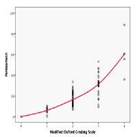
Correlation of levator ani muscle strength measurement between Modified Oxford Grading Scale and perineometer on pelvic organ prolapse patient

Published: 30 June 2021
Abstract Views: 2700
PDF: 289
Publisher's note
All claims expressed in this article are solely those of the authors and do not necessarily represent those of their affiliated organizations, or those of the publisher, the editors and the reviewers. Any product that may be evaluated in this article or claim that may be made by its manufacturer is not guaranteed or endorsed by the publisher.
All claims expressed in this article are solely those of the authors and do not necessarily represent those of their affiliated organizations, or those of the publisher, the editors and the reviewers. Any product that may be evaluated in this article or claim that may be made by its manufacturer is not guaranteed or endorsed by the publisher.
Similar Articles
- Fernandi Moegni, Andrew Yurius Christian, Levator hiatal ballooning prevalence in pelvic organ prolapse patients and its relation to levator ani muscle strength , Urogynaecologia: Vol. 34 No. 1 (2022)
- Maxime Marcelli, Gilles Karsenty, Jean-Philippe Estrade, Aubert Agostini, Ludovic Cravello, Gérard Serment, Marc Gamerre, Factors influencing sexual function in women with genital prolapse , Urogynaecologia: Vol. 25 No. 1 (2011)
- Khaled Refaat, Constanze Fischer-Hammadeh, Mohamad Eid Hammadeh, Overview of pelvic floor failure and associated problems , Urogynaecologia: Vol. 26 No. 1 (2012)
- Anggrainy Dwifitriana Kouwagam, Fernandi Moegni, Budi Iman Santoso, Suskhan Djusad, Surahman Hakim, Tyas Priyatini, Alfa Putri Meutia, The role of levatorplasty procedure in improving genital hiatus area and symptoms in pelvic organ prolapse with ballooning in Indonesia , Urogynaecologia: Vol. 36 No. 1 (2024)
- Kristina Crafoord, Jan Brynhildsen, Olof Hallböök, Preben Kjølhede, Pelvic organ prolapse and anorectal manometry: a prospective study , Urogynaecologia: Vol. 26 No. 1 (2012)
- Marie-Andrée Harvey, The size of the cervix and its relationship with age and parity , Urogynaecologia: Vol. 29 No. 1 (2016)
- Ali Mahmood, Prianka Gajula, The role of the urogynecologist with sphincteroplasty: a multidisciplinary approach to a very common, yet devastating problem , Urogynaecologia: Vol. 26 No. 1 (2012)
- Munir'deen A. Ijaiya, Hadijat O. Raji, Treatment of pelvic organ prolapse , Urogynaecologia: Vol. 26 No. 1 (2012)
- F. Bernasconi, V. Napolitano, M. Primicerio, D. Lijoi, E. Leone, F. Armitano, M. Luerti, G.C. Sugliano, D. Vitobello, D. Riva, D. Gregori, SUI AND TVT IUS ND TVT SECURE SYSTEM: A PROSPECTIVE OBSERVATIONAL MULTICENTRIC STUDY. MORBIDITY AND SHORT-TERM PERCENTAGES OF SUCCESS , Urogynaecologia: Vol. 23 No. 3 (2009)
- Chendrimada Madhu, Penelope Harber, David Holmes, Unexpected benefits and potential therapeutic opportunities of tension free vaginal tape for stress urinary incontinence , Urogynaecologia: Vol. 27 No. 1 (2013)
1-10 of 86
Next
You may also start an advanced similarity search for this article.

